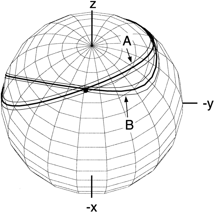
Figure 5.
Bands of allowed vector orientations for the Gln40 backbone amide 15N-1H bond vector in ubiquitin, in the frame of the crystal structure (1UBQ). Band A corresponds to the orientations compatible with the experimental dipolar coupling value measured in neutral bicelles. Band B corresponds to allowed orientations in bicelles charged with CTAB. The solid dot marks the orientation of the N-H vector when the proton is model-built into the crystal structure, assuming that HN is located on the line bisecting the C′-N-Cα angle. (Reprinted, with permission, from Ramirez and Bax 1998.)

