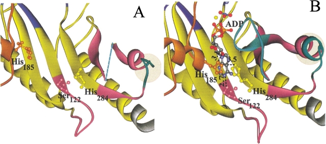
Figure 5.
Details of the (A) substrate-free and (B) transition state structures near the nucleotide-binding site. Domain 2 (orange) contributes His185, domain 3 (purple) contributes Ser122, and the fixed domain contributes His284, which all interact with the adenine ring of substrate ADP (B). Flexible loops 309–320 and 291–299 (cyan) are visible in the closed form of the enzyme (B), but residues 310–319 are disordered in the open form (dotted line in A showing connection). The extension of helix 16 (residues 296–299) in the closed structure is highlighted. Distances are shown in Å units.

