
Figure 3.
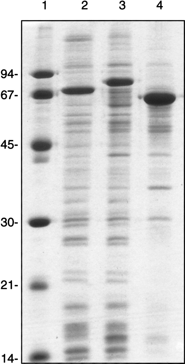
SDS-polyacrylamide gel electrophoresis of di-iron proteins expressed from pHB-MALc2X recombinants. Lane 1, molecular weight standards. Lane 2, membrane fraction from E. coli C43 (DE3) expressing the MBP-AtAOX fusion. Lane 3, membrane fraction from E. coli C43 (DE3) expressing the MBP-PTOX fusion. Lane 4, membrane fraction from E. coli JF496 expressing the MBP-PaCoq7 fusion. Two micrograms of protein loaded per lane. The expected mass of each fusion is MBP-AtAOX, 78.5 kD; MBP-PTOX, 85.7 kD, and MBP-PaCoq7, 68.8 kD.