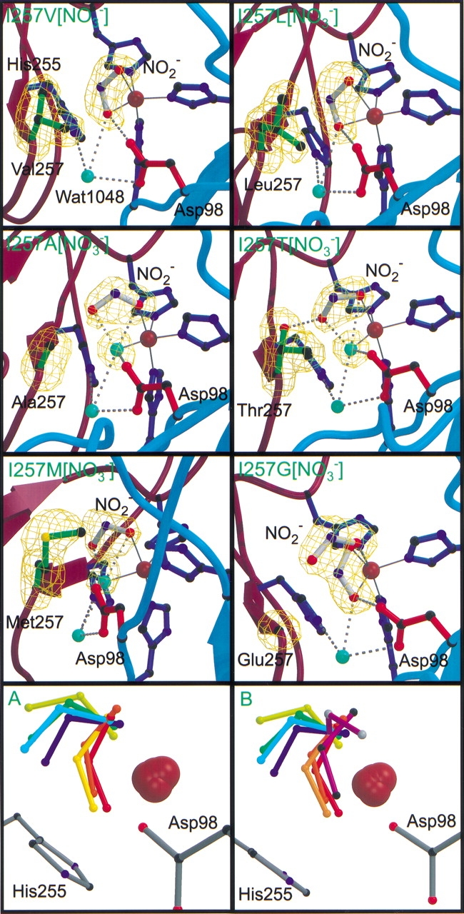
Figure 1.

Crystal structures of the six nitrite-soaked I257 variants in the upper six panels. Hydrogen bonds are shown as dashed gray lines with ligand bonds drawn in solid, dark gray lines. Water molecules are drawn as aquamarine spheres. Copper atoms are gray; nitrogen atoms are dark blue, oxygen atoms are red, and sulfur atoms are yellow. The backbone of monomers B and C are shown in burgundy and in teal, respectively. Bonds of the nitrite molecule bound in the active site are white. Omit Fo–Fc electron density maps are contoured at 4σ and drawn as a green wire mesh. Panels A and B depict the multiple conformations of the nitrite bound in the active site of the Ile257 variants and including nitrite bound to the oxidized D98N (black atoms) and H255N (gray atoms) crystal structures (Boulanger and Murphy 2001). With the exception of the purple nitrite molecules in panel B, nitrite molecules are blue to red with increasing specific activity: Red, I257V[NO2−]; orange, native NiR from Alcaligenes faecalis S-6; light orange, I257L[NO2−]; yellow, I257M[NO2−]; green, I257A[NO2−]; cyan, I257G[NO2−]; blue, I257T[NO2−].
