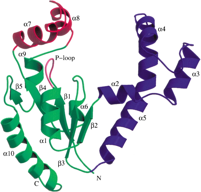
Figure 1.
Ribbon diagram depicting the E. coli DPCK monomer structure. The LID domain is red and the CoA-binding domain is blue, whereas the rest of the molecule is green. The nucleotide-binding P-loop motif is marked in pink. Secondary structural elements are marked. Figures 1 ▶, 2 ▶, and 4 ▶ were produced with Molscript (Kraulis 1991) and Raster3D (Merrit and Bacon 1997).

