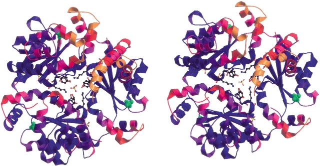
Figure 2.
Stereo ribbon diagram of the E. coli DPCK trimer structure, looking down the pseudo-threefold axis. The molecule is colored according to the temperature factors of the Cα atoms in each residue, from blue (B = 14 Å2) to orange (B = 60 Å2). Sulfate ions in the structure are displayed, as well as the side chains of residues Ser-119, Tyr-121, and Lys-122 from each monomer. Sulfates bound to the P-loop motif are shown as space-filling models in green.

