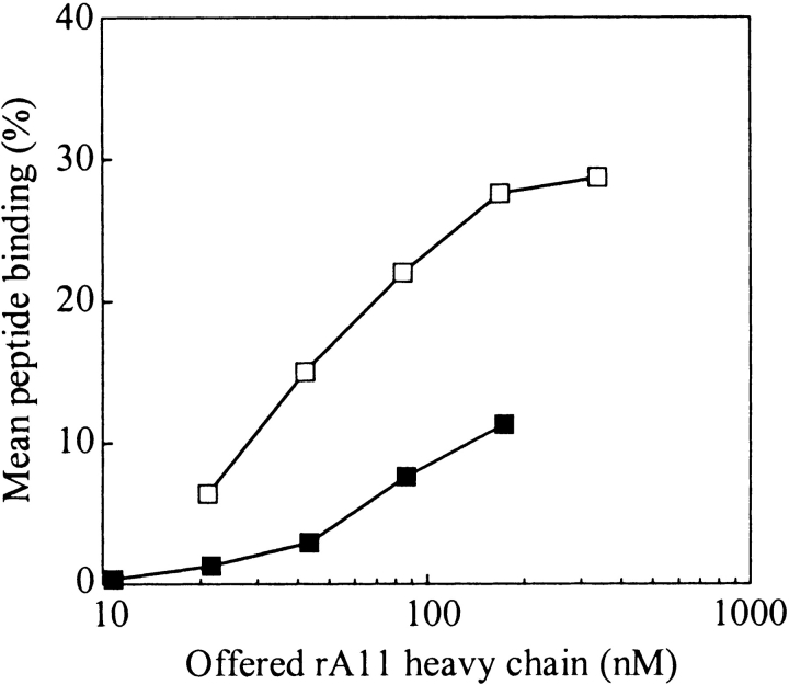
Figure 7.
Dose-response curve for purified rA11 isomer 0 (reduced) and isomer 1 (oxidized). Graded concentrations of purified A11 isomer 0 and 1 were diluted 100-fold into 100 mM Tris-maleate (pH 6.6) buffer containing human β2m (3 μM) and a specific radiolabeled peptide (15,000 cpm) and 1 mg/mL pluriol. The mean peptide binding values were calculated as described in Materials and Methods. Empty squares indicate rA11 isomer 1; solid squares, rA11 isomer 0. The standard deviation of duplicate peptide binding measurements was typically within 5%.

