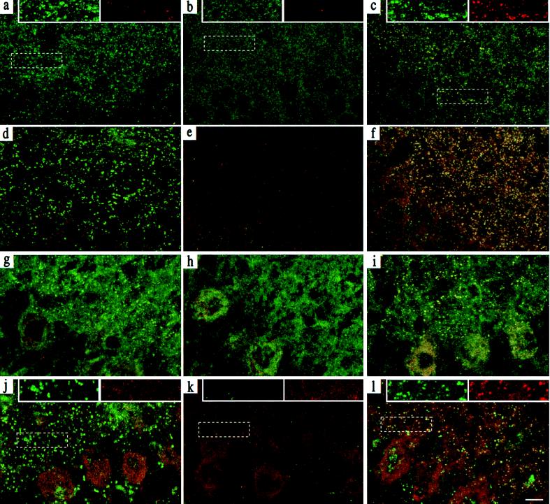
Figure 3.
Clustering of γ3 subunit-containing receptors and gephyrin in γ3tg/γ20/0 hippocampus and cerebellum. Parasagittal sections through the CA1 region of P14 hippocampus (a–f) (n = 4–6 per genotype) were stained with Abs specific for the α2 and γ3 subunits (a–c; α2 green, γ3 red) and for gephyrin and the γ3 subunit (d–f; gephyrin green, γ3 red) and visualized by confocal microscopy. The images were digitally superimposed to illustrate the presence or absence of colocalization between the markers used (yellow puncta). The punctate α2 (a) and gephyrin (d) staining, which is not colocalized with the scarce γ3 subunit IR in wt mice, was completely lost in γ20/0 mice (b and e) and largely restored in γ3tg/γ20/0 mice (c and f). In the latter, it was extensively colocalized with the γ3 subunit, as shown in yellow. Parasagittal sections through the molecular layer of P14 cerebellum (g–l) were stained for the α1 (green) and γ3 subunit (red; g–i) and for gephyrin (green) and the γ3 subunit (red; j–l). The punctate α1 subunit (g) and gephyrin (j) staining in wt brain was completely lost in γ20/0 (h and k) and largely restored in γ3tg/γ20/0 brain (i and l). Whereas specific γ3 subunit IR was absent in wt and γ20/0 cerebellum, it was readily detected and extensively colocalized with the α1 subunit in γ3tg/γ20/0 cerebellum. The data were reproduced with four to six animals per genotype. Insets at the top of panels a–c and j–l show enlargements of the boxed areas to illustrate the colocalization of clustered GABAA receptor and gephyrin IR in color-separated images. (Scale bar, 10 μm.)

