Figure 1.
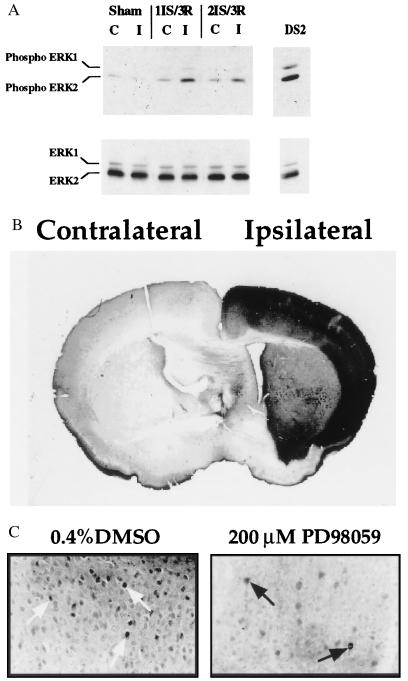
(A) Increase in ERK1/2 phosphorylation after ischemia and reperfusion. Western blot analysis was performed on lysates (40 μg per lane) from the contralateral (C) and ipsilateral/ischemic (I) side of the brain after 1 hr of ischemia (1IS) followed by 3 min of reperfusion (3R) or 2 hr of ischemia (2IS) followed by 3 min of reperfusion (3R). Western analysis was performed with phospho-specific ERK1/2 antibodies (New England Biolabs) (1:1000 dilution) (Upper). The blot was stripped and reprobed with C-14 ERK2-specific antibodies (Santa Cruz Biotechnology) (1:1000 dilution) (Lower). DS2 total cell lysate (40 μg) was used as a control for ERK1/2 phosphorylation. DS2 is a clonal cell line that exhibits constitutively active ERK1/2 (23). (B) Increased ERK1/2 phosphorylation in the cortical region of the brain after 1 hr of focal cerebral ischemia and 3 min of reperfusion. Immunohistochemistry of brain sections (40 μm thick) was performed with phospho-specific ERK1/2 antibodies. Contralateral (nonischemic) and ipsilateral (ischemic) sides of the brain are indicated. (×100.) (C) Pretreatment of mice with PD98059 leads to decreased ERK1/2 phosphorylation in the cortical region of the brain after 2 hr of focal cerebral ischemia and 3 min of reperfusion. Immunohistochemistry of brain sections (40 μm thick) was performed with the phospho-specific ERK1/2 antibodies. Nuclear staining is indicated by the arrows. (×400.) Dunnett’s posthoc tests. (C) PD98059 neuroprotection is maintained 3 days after ischemia. Brain sections were analyzed as described in the text. Numbers reflect the values from seven mice per treatment; ∗, P < 0.05. The data were analyzed by Student’s t test. Note: The number above each bar reflects the mean infarct volume as a percentage of the contralateral hemisphere to correct for edema, and was calculated using the following formula: (contralateral volume − ipsilateral undamaged volume) × 100/contralateral volume (8). (D) Pretreatment with SB203580, an inhibitor of p38 MAP kinase, is not neuroprotective. Mice were pretreated with 2 μl of 100 μM SB203580 or 0.2% DMSO 30 min before 2 hr of focal cerebral ischemia followed by 22 hr of reperfusion. The numbers reflect the values from four mice per treatment.

