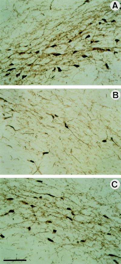
Figure 4.
Histological analysis of SN DA neurons of treated rats. Representative pictures of 14-μm-thick coronal sections through the SN processed for TH immunohistochemistry are shown. (A) Section contralateral to the lesion. (B and C) Sections ipsilateral to the lesion of animals injected with Ad-βGal (B) or Ad-GDNF (C). The aspect of the ipsilateral SN from rats that received only 6-OHDA is comparable to those of rats that received 6-OHDA and Ad-βGal (see B). (Bar = 100 μm.) The number of TH+ cell bodies and density of TH-stained fibers were both higher in rats that received Ad-GDNF than Ad-βGal (C versus B).

