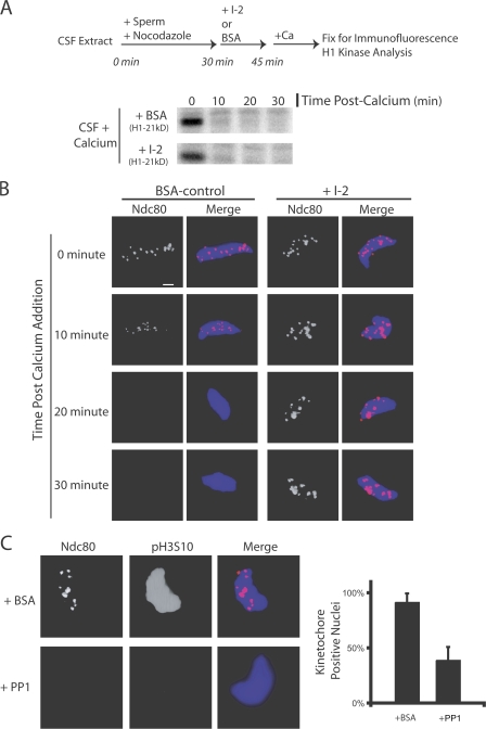
Figure 7.
PP1 is both necessary and sufficient to disassemble kinetochores on nuclei in extracts. (A and B) Kinetochores were assembled for 30 min in CSF extracts. I-2 or BSA was added to extracts and, 15 min later, the extract was driven out of M phase. (A) Both BSA- and I-2–treated extracts exited mitosis biochemically after the addition of calcium, as judged by histone H1 kinase activity. (B) In I-2–treated extracts, Ndc80 was retained at the kinetochore for 30 min after M phase exit. In control BSA-treated extracts, Ndc80 staining was reduced by 10 min and was undetectable by 20 min. (C) Nuclei with assembled kinetochores were isolated, washed, and mixed with purified PP1 enzyme. The addition of PP1 caused the dephosphorylation of H3S10 and the loss of kinetochore staining on the nuclei compared with BSA-treated nuclei. In control BSA-treated samples, 92% of nuclei stained positive for Ndc80. In PP1-treated samples, 39% of nuclei stained positive for Ndc80 (n > 100 nuclei per condition). Graphs show mean plus standard deviation. Bar, 5 μm.

