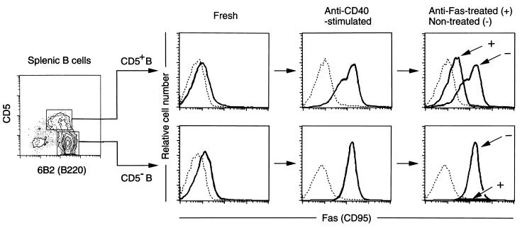
Figure 2.
Comparisons of Fas expression levels and sensitivity to Fas-mediated apoptosis between CD5+ and CD5− B cells. Splenic CD5+ and CD5− B cells from 2-month-old NZB/W F1 mice were FACS-sorted and stimulated with anti-CD40 mAb for 3 days, followed by incubation in the presence (Anti-Fas-treated) or absence (Non-treated) of anti-Fas mAb for 24 hr. Fas expression levels on PI-negative living cells were examined by three-color flow cytometry. Dotted line depicts autofluorescence.

