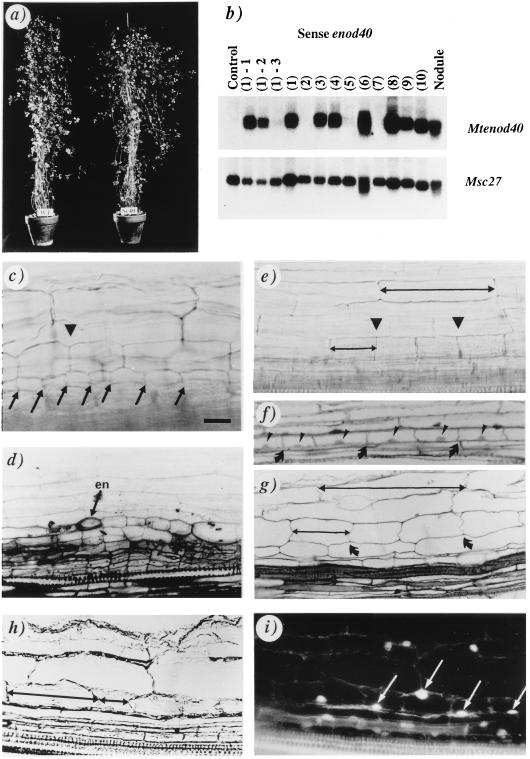
Figure 1.
Analysis of M. truncatula transgenic plants carrying the 35S–Mtenod40 construct. (a) An F2 plant (Se40) overexpressing Mtenod40 compared with a wild-type plant (WT). (b) Northern blot analysis of one control and 10 transgenic enod40 plants with an Mtenod40 probe. Progeny of the enod40 transgenic plant 1 [lanes (1)-1 to (1)-3] are also included. Msc27 was used as RNA-loading control (see ref. 7). (c and d) Lateral root primordia with dividing cortical cells (arrowhead) and divisions in the pericycle (arrows; en, endodermis). (e–i) Mtenod40-induced cortical cell division (e, arrowheads; f and g, arrows). Divisions of pericycle cells cannot be detected. A nucleus is present in each cell (f and i). Compare the size of undivided and divided cortical cells (double arrows in e, g, and h). Dividing cortical cells in a bright-field micrograph (h), showing fluorescent nuclei after 4,6-diamidino-2-phenylindole staining (arrows in i). Whole roots (c and e), 2-μm sections (d, f, and g), 14-μm sections (h and i). [Bar = 20 μm (c, e, h, and i) and 25 μm (d, f, and g).]

