Figure 1.
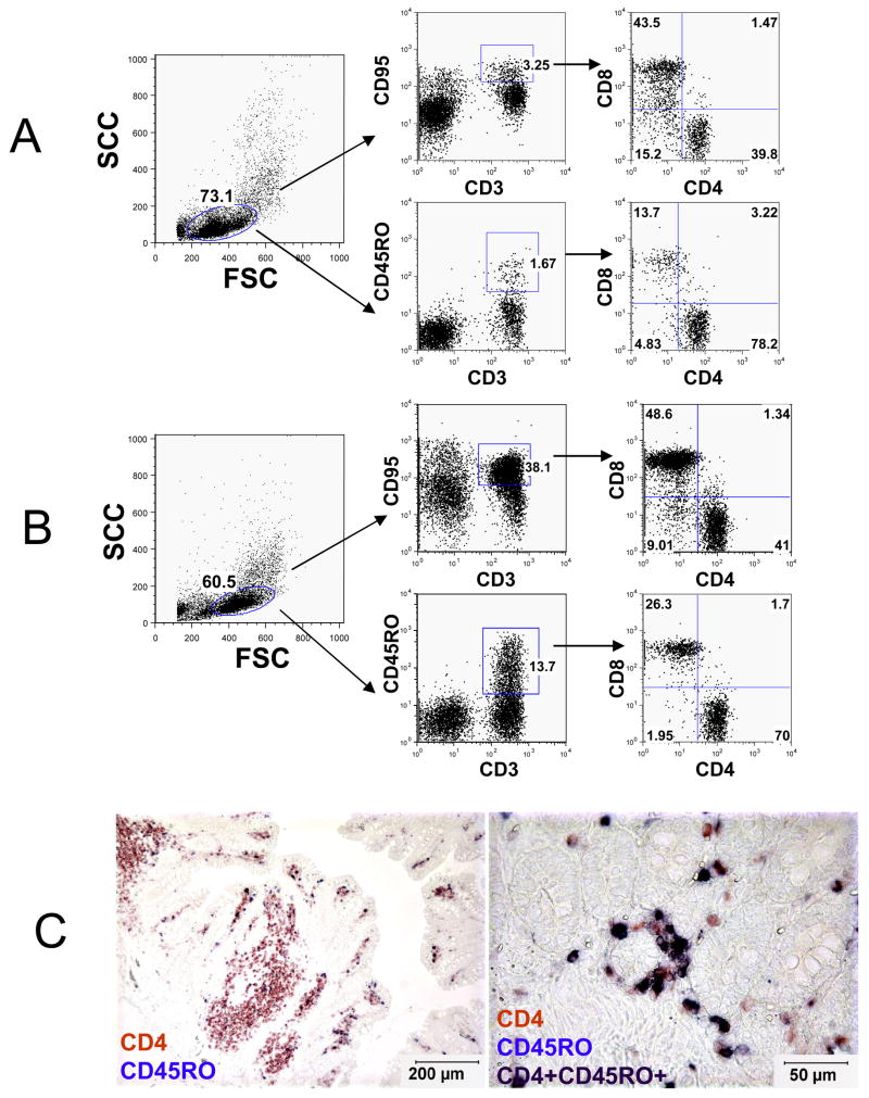
Flow cytometry dot plots (A, B) and dual immunohistochemistry staining (C) demonstrating that CD45RO (OPD4) is predominantly expressed on CD4+ T cells in rhesus macaques (A is from a 14 day old neonatal macaque (GL06) and B is from an adult 12 year old macaque (R533). Note that essentially all OPD4+ lymphocytes from both neonates (A) and adults (B) co-express CD3 in contrast to CD95, which also labels a subpopulation of non-CD3+ lymphocytes. The left panel indicates the lymphocyte gate and the center plots demonstrate CD45RO expression on CD3+ cells. The right plots indicate that the majority of OPD4+ (CD45RO) T cells are also CD4+ in both adult and neonatal macaques. C) Dual immunohistochemistry for CD4 (red) and CD45RO (blue) on paraffin-embedded intestinal sections showing CD45RO (OPD4) is expressed mostly on CD4+ T cells (purple/black, black arrows). The image on the right is a higher magnification of the intestine showing CD4+CD45RO+ cells as dark purple (black arrows) and a few CD4+ CD45RO neg cells in red (white arrows).

