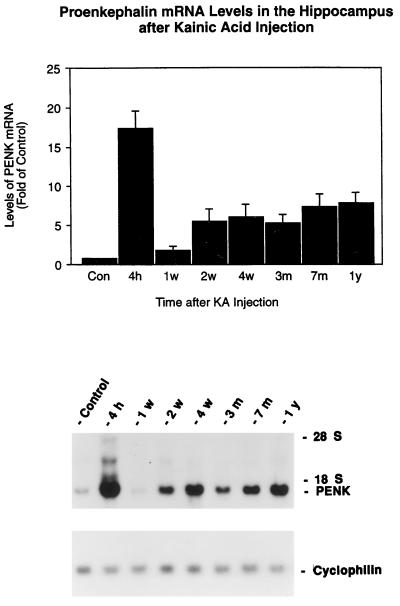
Figure 1.
Time course of the expression of PENK mRNA in the rat hippocampus after kainate injection. Rats were injected with kainate (7.25 mg/kg) s.c.. Hippocampi (n = 4) were collected at different time points and analyzed with Northern blots. (Upper) The autoradiographic signals corresponding to PENK and cyclophilin mRNA were quantified by laser densitometry. Ratios of PENK/cyclophilin mRNA density were calculated. Data represent means ± SEM. P < 0.01 at all time points compared with control except at 1 week (two-way analysis of variance, Bonferroni–Dunn post hoc tests were performed for between groups comparison). Control represents the mean of different time points from 1 week, 3 months, 7 months, and 1 year after a single injection of saline. There was no significant difference in the levels of PENK mRNA from these of control hippocampi. (Lower) Representative autoradiogragh of a Northern blot probed with 32P-labeled PENK and cyclophilin cRNAs showing the effects at 4 hr, 1 week, 2 weeks, 1 month, 3 months, 7 months, and 1 year after kainate treatment. The control sample was from the 3-month time point.

