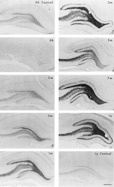
Figure 3.
Expression of ENK-IR in the rat hippocampus after kainate treatment. Representative immunocytochemical photomicrographs from three separate experiments are shown. Hippocampal immunostainings for ENK-IR from saline-treated rats at different time points were similar; photomicrographs from 6 hr (6 h) and 1 year (1 y) are shown. The kainate-treated groups include 6 hr (6 h), 2 weeks (2 w), 3 weeks (3 w), 1 month (1 m), 3 months (3 m), 7 months (7 m), and 1 year (1 y). Dramatic increases in ENK-IR were observed at 3 weeks after kainate treatment and persisted for up to 1 year. Note the progressive increase in the density of ENK-IR in the inner molecular layer of the dentate gyrus (sprouting of mossy fibers) in kainate-treated animals. (Scale bar = 400 μm.)

