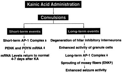
Figure 6.
Flow diagram of the biochemical and morphological changes in the hippocampus after treatment with kainate. Single systemic injections of kainate result in both short-term and long-term changes in the hippocampus. The short-term event is reflected by the rapid induction of AP-1 transcription factors, which may underlie the rapid induction of PENK and PDYN in the hippocampus. Elevated expression of the opioid peptides in the hippocampus lasted for a few days and returned to normal levels. This is the end of the short-term event. The long-term event after the kainate injection may ultimately lead to increases in the seizure susceptibility of the animals. Degeneration of the hilar inhibitory interneurons is the key event, which sets up the following events. Disinhibition of dentate granule cells increases changes of granule cell activity. This may activate the long-term AP-1 transcription factor. The loss of the targets of the mossy fibers will result in sprouting. These may induce the second phase of the increase of PENK expression in the granule cells. All of these events will result in increases of seizure activities.

