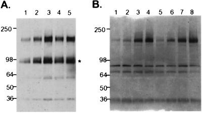
Figure 5.
Tyrosine phosphorylation of transfected and endogenous ErbB4 by NRG3EGF.Fc. (A) K562erbB4 cells were untreated (lane 1), or treated with 2.5 nM (lanes 2 and 4) and 25 nM (lanes 3 and 5) of NRG3EGF.Fc for 3 min (lanes 2 and 3) and 8 min (lanes 4 and 5). The cells were lysed and immunoprecipitated by anti-ErbB4 antibody, and probed with peroxidase-conjugated anti-phosphotyrosine antibody (Transduction Laboratories). *, a truncated ErbB4. The Mr (in kDa) is indicated at left. (B) MDA-MB-453 cells were untreated (lane 1) or treated with 2 nM, 20 nM, or 200 nM of NRG3EGF.Fc (lanes 2, 3, and 4, respectively), with 2 nM, 20 nM, or 200 nM of NRG3EGF.H6 (lanes 5, 6, and 7, respectively) or with 20 nM NRG1EGF (lane 8) for 3 min. The cells were lysed and subjected to immunoprecipitation and immunoblot as described in A. The Mr (in kDa) is indicated at left. For the blots shown in A and B, equal loading was confirmed by reprobing with anti-ErbB4 antibody (not shown).

