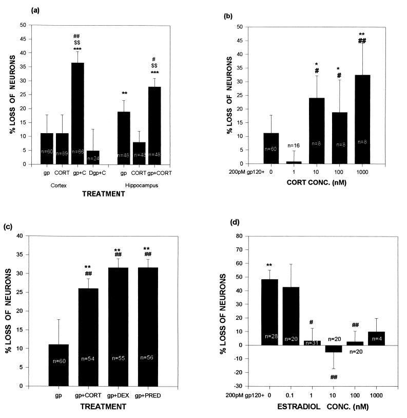
Figure 1.
Exposure to GCs exacerbates gp120-induced cell death in neuronal cultures, whereas estradiol is protective. Data are expressed as percentage of numbers of neurons in control wells (no gp120 or steroid) in the same plate. (A) In both hippocampal and cortical cultures, neuron loss was more pronounced in cultures treated with gp120 and 1 μM corticosterone than in cultures treated with either agent alone. (B) gp120-induced neuron loss varied with corticosterone concentrations. Significant levels of gp120-induced neurotoxicity coincided with corticosterone concentrations above the Kd (approximately 5 nM) of the type II GC receptor. (C) gp120-induced neuron death was worsened by the 1 μM concentrations of the synthetic GCs dexamethasone and prednisone. (D) Treatment of cortical cultures with estradiol reduced gp120-induced neurotoxicity. ∗∗ and ∗∗∗ indicate significant difference at the P < .01 and .001 levels, respectively, when compared with control (0% neuron loss, derived from cultures exposed to neither gp120 nor any steroid); # and ## indicate significant difference at the P < .05 and .01 levels, respectively, when compared with gp120 alone; $$ indicates significant difference at the P < .01 level, when compared with hormone alone; Newman–Keuls post-hoc tests after ANOVA. gp, gp120 (200 pM); CORT, corticosterone; Dgp, denatured gp120; DEX, dexamethasone; PRED, prednisone.

