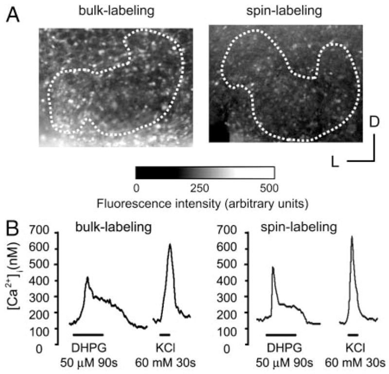
FIG. 1.

Comparisons between bulk-labeling and spin-labeling. A: examples of lateral superior olive (LSO) slices labeled using bulk (left) and spin-labeling (right) at postnatal day 0 (P0: the day of birth). Images show fluorescence at 360 nm (exposure time 20 ms, ×20 objective). Dotted lines outline the boundaries of the LSO. D, dorsal; L, lateral. B: example of changes in intracellular Ca2+ elicited by bath application of (S)-3,5-dihydroxyphenylglycine (DHPG, 50 μM, 90 s) and KCl (60 mM, 30 s). LSO neurons from a P3-old mouse.
