
Figure 5.
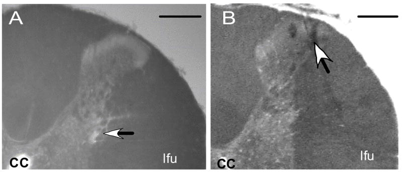
Representative examples of the dorsal right quadrant of coronal sections through the spinal cord illustrating the histological localization of the position (arrow) of the micropipette tip targeting (A) the intermediolateral cell column and (B) the dorsal horn within the fourth thoracic segment. Scale bars = 250 μm. cc, central canal; lfu, lateral funiculus