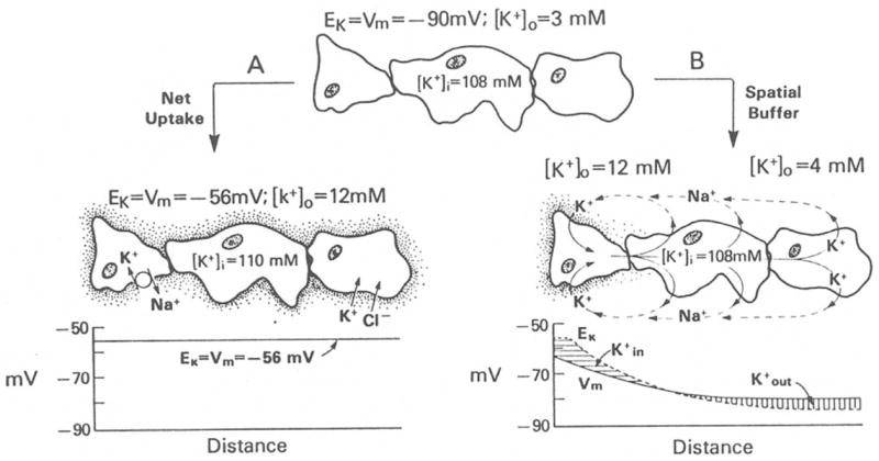
Fig. 2.

Diagram depicting the role of glial cells in [K+]o homeostasis. Top: Glial cells are electrically coupled via gap junctions forming a functional syncytium. With [K+]o equaling 3 mM, the glial syncytium has a membrane potential of approximately −90 mV. (A) Net K+ uptake mechanism. When [K+]o is increased, glial cells accumulate K+ either by the activity of a Na,K–ATPase or by a pathway in which K+ is cotranported with Cl−. In this mechanism of [K+]o regulation, the membrane potential in the glial syncytium is spatially uniform at −56 mV. (B) K+ spatial buffering mechanism. Local increases of [K+]o produce a glial depolarization that spreads through the glial syncytium. The local difference in Vm and EKdrives the K+ uptake in regions of elevated [K+]o and K+ outflow at distant regions. The intracellular currents are carried by K+ and extracellular currents are mediated by other ions such as Na+. [From Orkand (1986), with permission.]
