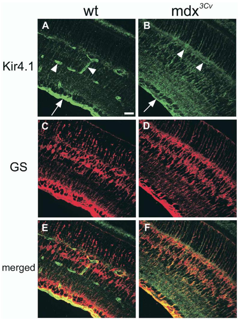
Fig. 6.

Kir4.1 channel localization in wild type and mdx3Cv (dystrophin knockout) mouse retinal sections. (A) Kir4.1 is concentrated at the inner limiting membrane (arrow) and to processes around blood vessels (arrowheads) in wild-type retina. (B) In the mdx3Cv mouse, Kir4.1 is more evenly distributed throughout the retina with a reduction in staining at the inner limiting membrane (arrow) and no apparent enrichment of Kir4.1 around blood vessels (arrowheads). The Müller-specific marker glutamine synthetase (C, D), and merged images (E, F) suggest the localization of Kir4.1 to Müller cells. Scale bar = 25 μm (A). [From Connors and Kofuji (2002), with permission.]
