
Figure 5.
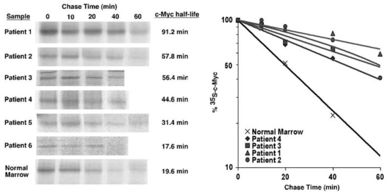
c-Myc protein half-life is prolonged in bone marrow samples from pediatric ALL patients. Stored diagnostic bone marrow samples from pediatric ALL patients were obtained after consent was received from the Oregon Health & Science University Institutional Review Board. Ficolled samples were thawed and maintained in media with 20% FBS for 16–20 h before analysis. Pulse-chase analyses were performed as described for Figure 2 and in Materials and methods. This figure shows results from the six samples in which sufficient c-Myc was detected for quantitation and assessment of half-life. Pulse-chase analyses were also performed on normal bone marrow samples as a control. The result for the ‘normal marrow’ sample shown is representative of three samples tested. c-Myc protein half-life is shown for each sample. The rate of c-Myc degradation over time is depicted in the graph for four samples with prolonged c-Myc half-life and for the representative normal marrow sample.