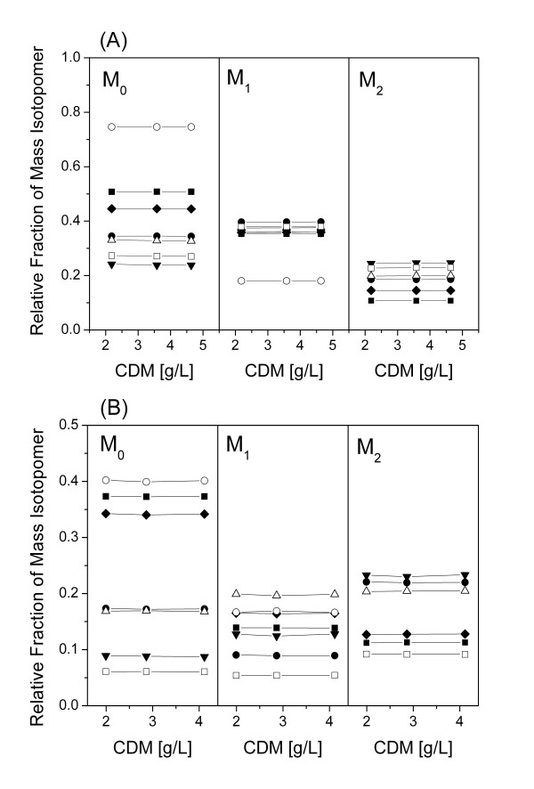
Figure 3.
Verification of isotopic steady-state during 13C tracer studies of C. glutamicum lysCfbr Δpyk grown on [1-13C] glucose (A) and an equimolar mixture of [13C6] glucose and naturally labelled glucose (B). The labelling patterns of the amino acids were determined from protein hydrolysates harvested at different cell dryx mass (CDM) concentrations during the cultivation. The amino acids shown here exemplarily stem form different parts of the metabolic network and comprise alanine (solid square), phenylalanine (open square), valine (closed circle), glycine (open circle), glutamate (closed triangle), threonine (open triangle) and serine (closed diamond). M0 (non labelled), M1 (single labelled) and M2 (double labelled) denote the relative fractions of the corresponding mass isotopomers.

