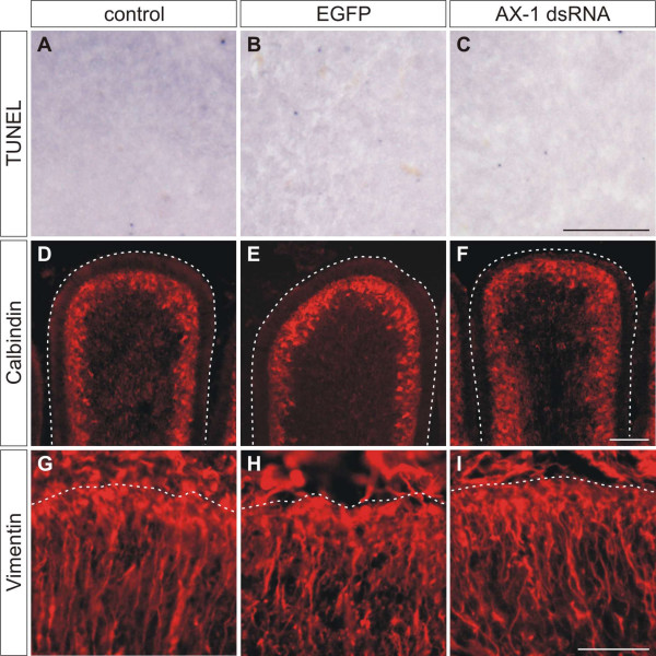
Figure 2.
Ex ovo electroporation and RNAi using long dsRNA does not induce apoptosis and does not disturb cerebellar organization.(a-c) Apoptosis was investigated by TUNEL in 30-μm-thick sagittal sections at HH35, one day after electroporation. Corresponding areas are shown in (a-c). No difference in apoptosis was detected between untreated embryos (a), embryos injected with EGFP plasmids alone (b), and embryos injected with AX-1 dsRNA (c). (d-f) The organization and the development of Purkinje cells analyzed by Calbindin staining at HH38 did not differ between untreated control embryos (d), EGFP-expressing control embryos (e), and embryos electroporated with AX-1 dsRNA (f). (g-i) Similarly, no changes in Bergmann glia cells were detectable at HH37 after staining for Vimentin, when untreated (g), EGFP-expressing control embryos (h), and embryos lacking AX-1 (i) were compared. Bar: 100 μm in (a-c), 200 μm in (d-f), and 50 μm in (g-i).

