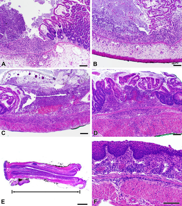
Figure 4.
Histologic changes during acute and chronic DSS colitis. Colon tissues from both control (A) and LMP-420-treated (B, 15 mg/kg LMP-420 i.p.) mice with acute DSS colitis demonstrated similar amounts of edema, acute inflammatory infiltrates, and focal ulceration. Chronic colitis generated by 3 cycles of 5 days of 3% DSS in drinking water, followed by 16 days of plain water resulted in development of severe chronic colitis (C) that was not altered by a 16 day treatment with treatment LMP-420 (D, 45 mg/kg i.p.). The cecum is shown in panels A and B and mid-colon is shown in panels C and D. Wild-type and RAG-2-/- mice with chronic DSS colitis developed extensive squamous metaplasia of the rectum (E, F) that in some cases extended proximally for > 1 cm. The bar equals 100 μm except for panel E, where bar = 1 mm.

