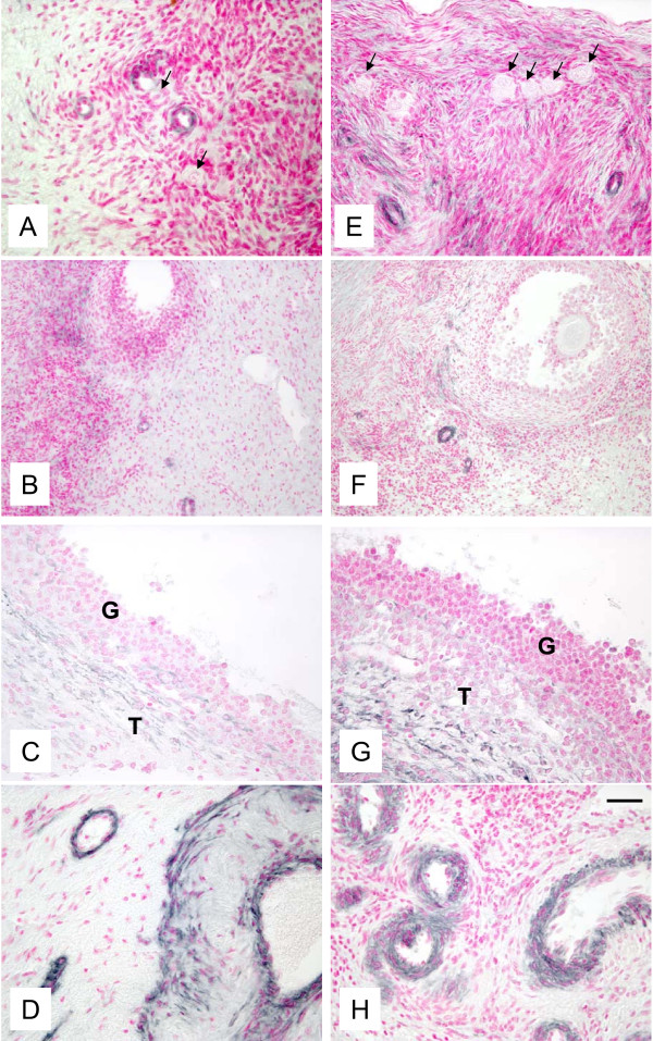
Figure 6.
Representative micrograph of staining for VEGF (black color) in ovarian tissues from autotransplanted (A, B, C, D; left column) and control (E, F, G, H; right column) ewes. Note presence of VEGF in blood vessels in the area containing primordial/primary and antral follicles (A, B, E, F), in the theca layer of preovulatory follicles (C, G), and in the large blood vessels in the ovarian stroma (D, H). Arrows indicate primordial/primary follicles. Control sections did not exhibit any positive staining (see Fig. 8 insert). Bar = 50 μm for A, B, D, E, G, H and 100 μm for B and F.

