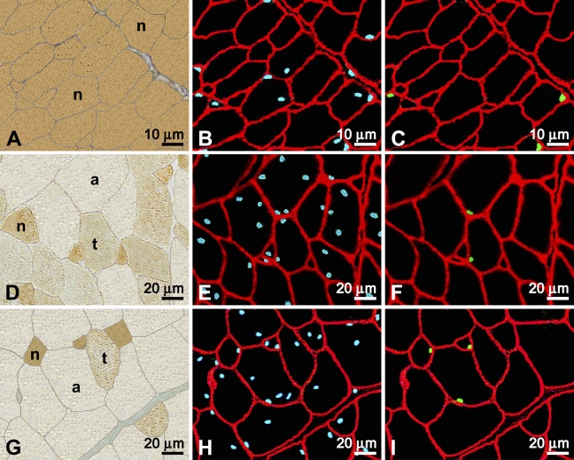
Figure 1.
Immunocytochemical labeling of serial cross-sections of developing chicken pectoralis. Muscles were from birds aged 9 (A–C), 49 (D–F), and 79 (G–I) days posthatch. (A,D,G) Viewed by brightfield and labeled by the anti-neonatal MyHC antibody to distinguish the type of fiber profile (neonatal, n; transforming, t; adult, a). (B,E,H) Viewed under epifluorescence and sections are cut serially, respectively, to A,D,G. All nuclei appear blue due to labeling by Hoescht, and basal laminae appear red with labeling by anti-laminin. (C,F,I) Same sections as B,E,H, respectively, showing satellite cell (SC) nuclei in green labeled by anti-Pax7 and the basal laminae in red with anti-laminin.

