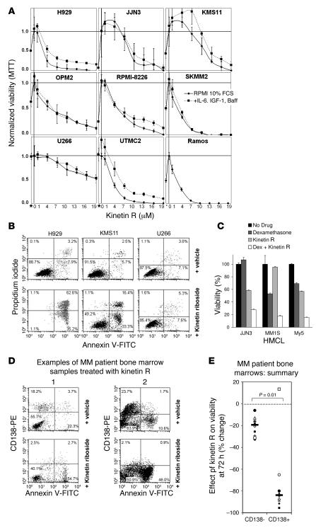
Figure 5. Kinetin riboside induces growth arrest and apoptosis in HMCL and primary myeloma cells and shows synergistic cytotoxicity with corticosteroids.
(A) HMCL viability following kinetin riboside (1–20 μM) in the presence or absence of MM growth cytokines IL-6 (10 ng/ml), IGF-1 (100 ng/ml), and BAFF (25 ng/ml) by MTT assay at 72 hours. The Burkitt lymphoma line RAMOS is also shown. *MTT on day 0 without drug. (B) Induction of apoptosis in HMCL by vehicle or kinetin riboside at 96 hours, assessed by annexin V and propidium iodide uptake. (C) Synergistic cytotoxicity induced by kinetin riboside (5 μM) and dexamethasone (10 nM) in JJN3, MM1S, and MY5 HMCL, assessed by MTT assay at 48 hours; graph shows the mean of triplicates ± SEM. (D) Two examples of flow cytometric analyses of MM patient bone marrow samples treated with vehicle or kinetin riboside (10 μM), showing preferential loss of viable primary CD138+ tumor cells (left upper quadrants) versus CD138– hematopoietic cells (left lower quadrants) with kinetin riboside at 72 hours. Following kinetin riboside treatment, CD138+ plasma cells became annexin V positive and CD138 negative, consistent with apoptosis. (E) Effects of kinetin riboside on CD138+ and CD138– compartments in 10 MM patient bone marrow samples at 72 hours. CD138+ and CD138– viability responses are compared by Mann-Whitney test. CD138+ (primary tumor) cells are preferentially killed.

