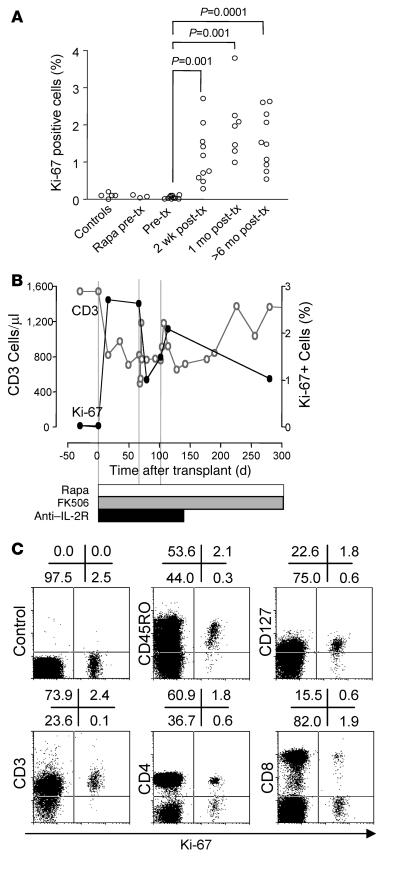
Figure 2. Detection and characterization of in vivo proliferating T cells following transplantation.
(A) Percentage of fixed peripheral blood lymphocytes expressing Ki-67, a nuclear marker of ongoing proliferation, in 5 normal nondiabetic subjects (controls), 3 patients with type 1 diabetes who received rapamycin (Rapa) as monotherapy prior to islet transplantation (tx), and 10 patients with type 1 diabetes before and after islet transplantation. In 2 of these patients, immunosuppression was stopped as a result of graft failure. Significant increases between medians are indicated. (B) A single representative case (from 10 studied) of sequential CD3+ T cell counts and the frequency of Ki-67+ lymphocytes after islet transplantation. (C) Phenotype of peripheral blood Ki-67+ cells in representative patient hSR-056-ITA-Ed07 following islet transplantation. Cells were stained for Ki-67 together with an isotype control, CD45RO, the IL-7 receptor CD127, CD3, CD4, or CD8. Numbers denote percentage of cells in the respective quadrants.

