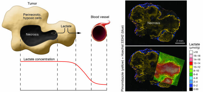
Figure 1. Mechanisms underlying increased lactate concentrations in hypoxic and necrotic versus normoxic, well-perfused tumor areas.
Necrosis occurs within the hypoxic core of the (experimental) tumor. Lactate produced by perinecrotic, hypoxic cells is cleared through the microvasculature. Lacking a route of drainage, lactate accumulates in the necrotic cavity. In addition, hypoxia leads to an increase in baseline lactate production rate via the Pasteur effect (16). Consequently, the concentration of lactate in hypoxic tumor areas is determined by oncogenic/hypoxic lactate production, lactate backflow from necrosis, and other factors, such as vascular efficiency and substrate availability. The right panel illustrates these principles in a human cervical carcinoma (SiHa) xenografted in a mouse. The administered hypoxia marker pimonidazole (yellow) labels hypoxic cells, whereas external Hoechst 33342 (blue) marks better-perfused and oxygenated parts of the tumor. Lactate was determined from cryoslices using quantitative bioluminescence microscopy. The images were color encoded and coregistered with the pimonidazole/Hoechst image.

