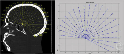
Figure 1.
The mid-sagittal ray analysis, precursor to 3DVA. (A and B) Analysis is performed in the mid-sagittal plane only. A set of radial vectors at 10° increments eminates from the posterior ledge of the sella. Measurements are taken to the outer table. This analysis localizes regional differences (like frontal vs occipital bossing in scaphocephaly) but is limited to 2 dimensions.

