
Figure 4.
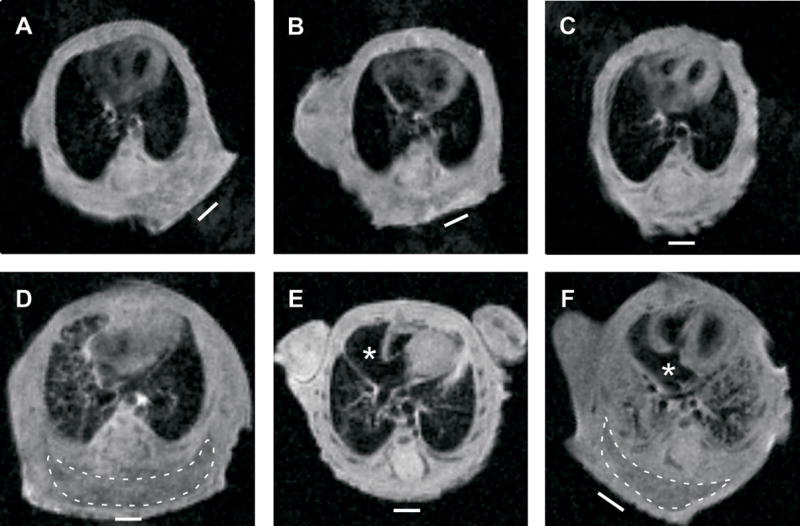
Magnetic resonance imaging. MRI imaging on wild type (n = 8) and Tpst DKO (n = 4) was performed on pups delivered by Cesarean section at E19.5 as described in Materials and Methods. Coronal images at the level of the entry of the inferior vena cava into the right atrium from three wild type (panels A–C) and three Tpst DKO (panels D–F) pups are shown. Images from the fourth Tpst DKO pup are not shown but look nearly identical to the image in panel E. The right atria are marked by an asterisk and areas of dependent edema are surrounded by a dashed line. The white bar indicates the horizontal plane during the imaging sequence.