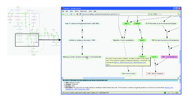
Figure 7.
Visualization of an E. coli pathogenesis network in Phinet. A click on each node provides detailed information about a biological object in the bottom frame. When a mouse cursor moves over a node, a brief description of the biological object will appear. An interaction between biological objects is represented by a centered gray ball and arrows between nodes. Once the centered gray ball is clicked, details about the specific interaction appear in the bottom frame. Subcellular locations of biological objects are differentiated by the node border colors. The biological object types (for example, protein or gene) are represented by a combination of the node background colors and shapes. The program also displays different interactions, such as inhibition (solid T sign), activation (solid arrow), and indirect effects (dashed line).

