
Figure 1. Ngb protein expression and cell viability in PC12 cells after transfection of Ngb siRNA and treatment of H2O2.
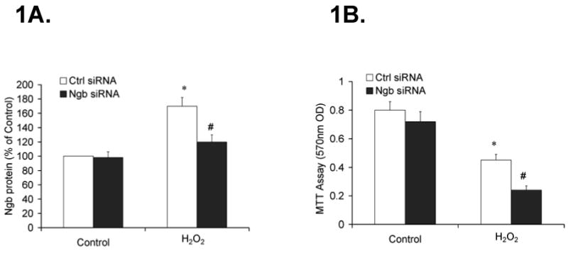
PC12 cells were transfected with Ng siRNA or control siRNA for 48 h. Following transfection, the cells were treated with H2O2 (0.1mM) or same volume of H2O (control) for 6 h. Ngb protein expression was determined by quantitative western blotting. Cell viability was assessed by MTT assay. 1A, Ngb protein expression following transfection of Ngb siRNA and treatment of H2O2. Data were expressed as a percentage of control siRNA in control group (n=6/group, *P<0.01 vs. control, #P<0.01 vs. control siRNA). 1B. Cell viability following transfection of Ngb siRNA and treatment of H2O2. Data were expressed as absolute OD values (570 nm OD) (n=6/group, *P<0.01 vs. control, #P<0.01 vs. control siRNA).