Figure 1.
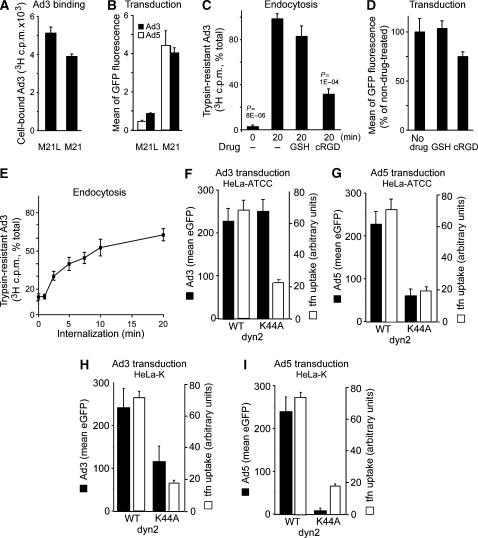
Infectious Ad3 endocytosis of HeLa cells requires αv integrins and to a low extent dynamin. (A) Human melanoma M21 or M21L cells were incubated with [3H]thymidine-labelled Ad3 in the cold and analysed for cell-associated radioactivity (106 cells, 0.75 μg Ad3). (B) M21 or M21L cells were transduced with Ad3-eGFP or Ad5-eGFP (MOI 5) and analysed by flow cytometry 8 h p.i. (C, D) [3H]thymidine-labelled Ad3 (8 × 10⁵/ml; 50 000 c.p.m.) was cold bound to HeLa-ATCC cells (2000 c.p.m. bound) and warmed for 0 or 20 min in the presence or absence of glutathione (GSH) or cyclic RGD peptide (cRGD, 0.2 mM), trypsinized in the cold, and analysed for cell-associated (internalized) or released [3H]thymidine-labelled Ad3 by liquid scintillation counting. Cells were treated with GSH or cRGD, infected with Ad3-eGFP (MOI 5), and analysed for GFP expression by flow cytometry 6 h p.i. (E) Kinetics of Ad3 endocytosis measured by trypsin resistance as described in panel C (100% equivalent to 2000 c.p.m.). Means of triplicate dishes of one representative experiment are shown (A–E). (F–I) Ad3-eGFP and Ad5-eGFP transduction (6 h) and transferrin–Alexa647 internalization (10 μg/ml transferrin in the last 30 min of infection, open bars) in HeLa-ATCC or HeLa-K cells transfected with WT dyn2 or K44A dyn2 for 48 h. Single-cell analysis by confocal microscopy (MOI 5), showing the mean of at least 40 blindly selected cells, is shown. One representative experiment is shown (F–I).

