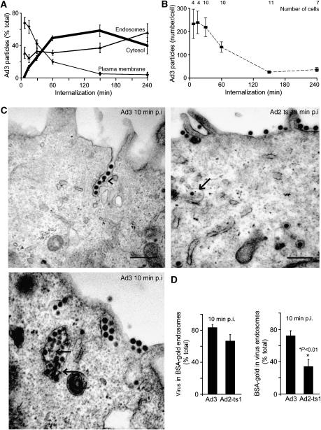
Figure 2.
Quantitative EM analyses of Ad3 endocytosis and endosomal escape. (A) Distribution of cold-bound Ad3 (5 × 10⁵ viral particles per cell, 4°C, 60 min) on the plasma membrane, endosomes, and the cytosol (bold line) upon internalization at 37°C. (B) Analyses of the total number of particles and cells. (C, D) Enrichment of Ad3 in fluid-phase-positive endosomes. HeLa cells were incubated with Ad3 or Ad2-ts1 in the cold (MOI as in (A)), washed, pulsed with BSA-gold for 10 min, fixed for ultrathin-section EM analyses, and quantified for viral particles in either gold-positive endosomes (arrows) or gold particles in endosomes that contain Ad3 or Ad2-ts1, respectively. Viruses in plasma membrane invaginations are indicated by an arrowhead.

