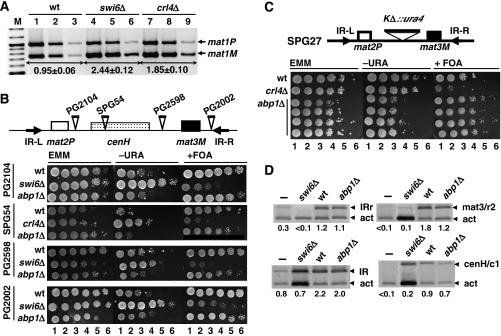
Figure 5.
Heterochromatin organisation of the mating-type region is not disrupted in abp1Δ cells. (A) Quantitative multiplex PCR determination of the predominant mating type adopted by wild-type (lanes 1–3), swi6Δ (lanes 4–6) and crl4Δ cells (lanes 7–9). PCR reactions were performed as in Figure 1D. The M/P ratios (±s.d.) are indicated below the corresponding lanes. (B) The effect of deleting abp1 on heterochromatin-mediated silencing was determined in several strains carrying ura4+ insertions at different positions of the silent domain of the mating-type locus. Sites of insertion are schematically indicated on top. Exponentially growing cells were plated as serial 10-fold dilutions (lanes 1–6) onto selective media with (panels EMM) or without uracil (panels −URA), or in the presence of FOA (panels +FOA). For each strain, several independent abp1Δ isolates were analysed and they all showed very similar results. (C) Similar results as in (B) but performed with strain SPG27, where the entire cenH element was replaced by ura4+. In this case, the results obtained with four independent abp1Δ isolates are presented. (D) Swi6 deposition at the silent domain of the mating-type region was determined in wild-type (lanes wt) and abp1Δ cells (lanes abp1Δ) by ChIP analysis using αSwi6 antibodies. Immunoprecipitated material was analysed as in Figure 3 with primers to amplify specific bands of the IR-R and IR-L regions (bands IRr and IR), the cenH element (bands cenH/c1) and a region just left of mat3 (bands mat3/r2) (see Supplementary Table SII for a description of the primers used). Lanes swi6Δ correspond to immunoprecipitation experiments performed with αSwi6 antibodies in a swi6Δ strain, missing Swi6. Lanes – correspond to mock immunoprecipitation experiments in which no antibodies were added. Numbers below each lane correspond to the ratio of the corresponding mating-type-specific band with respect to the control act band.

