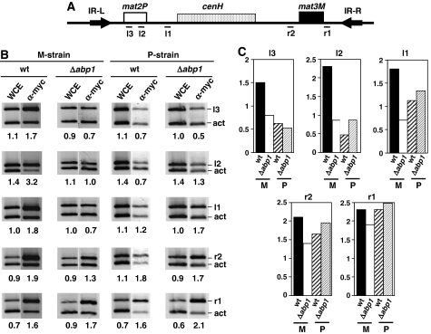
Figure 7.
Deletion of abp1 impairs spreading of Swi2–Swi5 to mat2. The effects of deleting abp1 on the distribution of Swi2–myc along the silent domain of the mating-type locus were determined in both a stable M-strain (mat1smto) and a stable P-strain (mat1PΔ17) by ChIP analysis using α-myc antibodies. (A) Regions of the silent domain of the mating-type locus where the presence of Swi2–myc was analysed (see Supplementary Table SII for a description of the primers used). (B) ChIP analysis corresponding to wild-type (wt) and abp1Δ cells derived from either a stable M-strain (panel M-strain) or a stable P-strain (panel P-strain). Material obtained after immunoprecipitation with α-myc antibodies was analysed as described in Figure 3 using primers specific for the indicated regions (bands r1, r2, l1, l2 and l3). Lanes WCE correspond to PCR products obtained from the input material before immunoprecipitation. Lanes α-myc correspond to the products obtained from the immunoprecipitated material. Numbers below each lane correspond to the ratio of the corresponding mating-type-specific band with respect to the control act band. (C) Quantitative analysis of the results shown in (B). For each of the mating-type-specific regions analysed, the relative fold of enrichment of the corresponding band in the immunoprecipitated material with respect to the input material (WCE) is presented for both wild type (wt) and abp1Δ cells in a stable M or P background.

