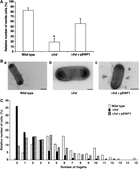
Figure 3.
Effects of frd and sdh deletions on swimming, assembly of flagella, and switching the direction of flagellar rotation. (A) Percentage of motile cells. Swimming cells were video-recorded and the fraction of motile cells was determined blindly. Values shown are the mean±s.e.m. of three experiments, 100–200 cells for each strain in each experiment. The asterisk indicates a statistically significant difference from the other columns (P<0.01 and P<0.05 for wild-type and Δfrd+pEWF1 columns, respectively; one-way ANOVA plus Tukey–Kramer tests). (B) Typical micrographs of strains RP437 (a; bar=1 μm), RP437Δfrd (b; 0.5 μm), and RP437Δfrd containing pEWF1 (c; 1 μm). (C) Distribution of the number of flagella per cell. Cells were negatively stained with uranyl acetate and photographed using a transmission electron microscope. The number of flagella of ∼200 cells of each strain were counted without prior knowledge of the strain being counted. The difference in the number of flagella between the strains was very significant (P<0.001; Krusal–Wallis test). Open columns, strain RP437; solid columns, RP437Δfrd; hatched columns, RP437Δfrd containing pEWF1.

