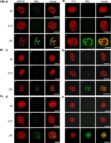
Figure 2. Endogenous SAP30 and YY1 Colocalize with NSs Filaments in the Nuclei of ZH Infected Cells.
Colocalization of endogenous SAP30 (A, C, and E) and YY1 proteins (B, D, and F) with NSs filament was analyzed by confocal microscopy in L929 wt330 cells uninfected (NI) or infected with C13 or ZH at m.o.i. 5 collected at 18 h p.i. (A and B), 5h (C and D) or 7 h p.i. (E and F). Each row represents a single optical section of the same nucleus. A, C, and E) Left panels (a, d, g) correspond to SAP30 distribution revealed with goat polyclonal anti-SAP30 antibodies. Middle panels (b, e, h) show subnuclear NSs distribution detected with anti-NSs rabbit polyclonal antibodies. Merged images of SAP30 and NSs are shown on right panels (c, f, i). (B, D, and F) Left panels (a, d, g) correspond to YY1 distribution revealed with mouse monoclonal anti-YY1 antibody. Middle panels (b, e, h) show subnuclear NSs distribution detected with anti-NSs rabbit polyclonal antibody. Merged images of YY1 and NSs are shown on right panels (c, f, i). Scale bar, 10 μm.

