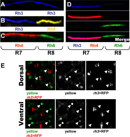
Figure 2. rh3 and rh4 Are Co-Expressed in yR7 Cells.
(A–D) Antibody staining of wild-type dissociated ommatidia. (A) DRA subtype: Rh3 (blue) is present in both R7 and R8. (B) p subtype: Pairing between Rh3 (blue) in R7 and Rh5 (yellow) in R8 is observed. (C) y subtype: Pairing between Rh4 (red) in R7 and Rh6 (green) in R8 is observed. (D) In the “dorsal” y subtype, Rh3 (blue) and Rh4 (red) are present in the same (pink) R7 and they are coupled with Rh6 (green) in R8.
(E) Visualization of the “yellow” pigment (green) and rh3>RFP (red) using cornea neutralization technique of dorsal and ventral regions of the eye. The “yellow” pigment can be visualized due to its inherent fluorescence in the green channel. Arrowheads point to yR7 rhabdomeres. Note that, contrary to “yellow,” RFP is not restricted to the rhabdomere, allowing the whole cell to be visualized. Residual signal in pigment cells surrounding each ommatidium is also observed in the green channel.

