
Figure 2. Deletion of miR-106a~363 or miR-106b~25 results in viable and fertile mice.
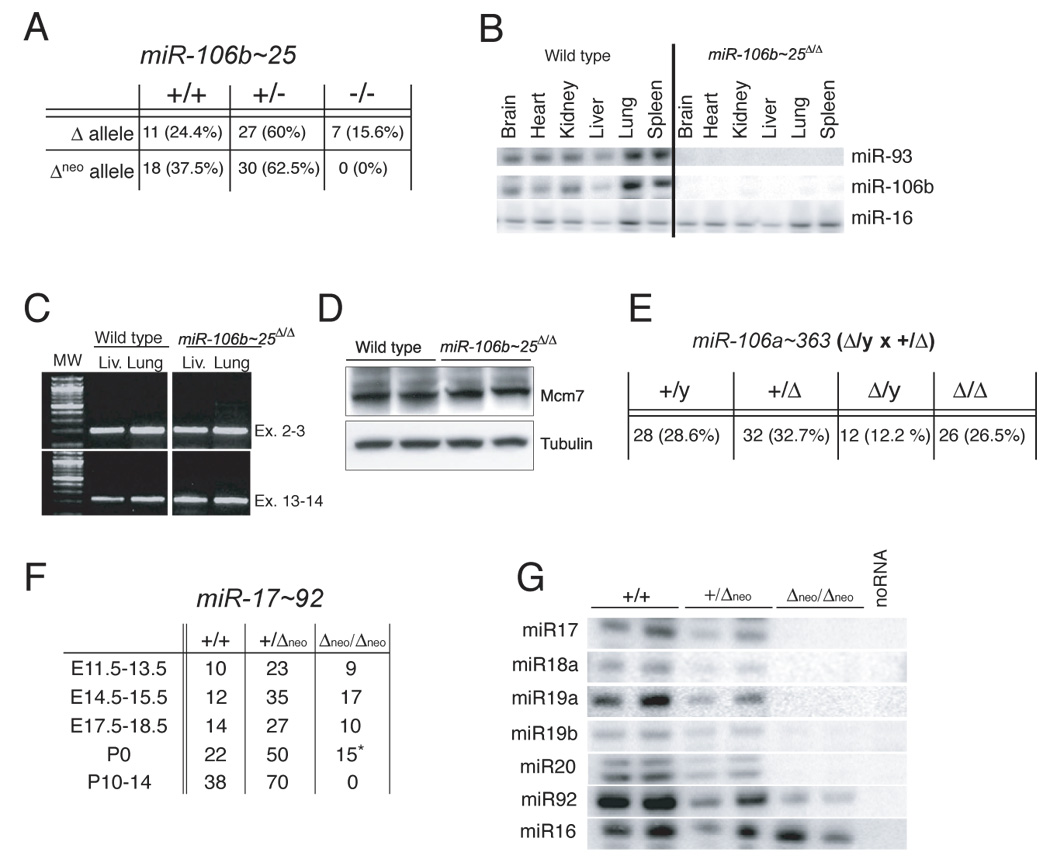
(A) Genotypes of mice from miR-106b~25+/Δneo and miR-106b~25+/Δ intercrosses. (B) RPAs on total RNA extracted from wild-type and miR-106b~25Δ/Δ adult tissues. (C) RT-PCR on RNA extracted from miR-106b~25Δ/Δ and wild-type lungs and livers. Primers designed to amplify the junctions of exons 13–14 were used to determine whether the deletion of miR-106b~25 from intron 13 affects splicing between these two exons. Amplification of the junction between exons 2 and 3 was used as control. (D) Mcm-7 Western blot on whole cell lysates from wild type and miR-106b~25 Δneo/Δneo mouse embryo fibroblasts. (E) Genotypes of mice from miR-106a~363Δ/Y × miR-106a~363+/Δ crosses. (F) Genotypes of mice from miR-17~92+/Δneo intercrosses determined at the indicated gestation stages. The asterisk indicates that all miR-17~92Δneo/Δneo newborn mice died soon after birth. (G) RPAs on total RNA from E13.5 embryos with the indicated genotypes.