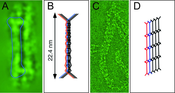
Figure 4.
DivIVA oligomers form a two-dimensional network as judged from cryonegative stain transmission electron microscopy images. A) Freshly purified DivIVA appears as a "doggy-bone" shaped particle. B) A tentative model for the hexameric DivIVA oligomer. C) Further oligomerization of DivIVA "doggy-bones" leads to two-dimensional network formation. D) A tentative model for the two-dimensional DivIVA network [100].

