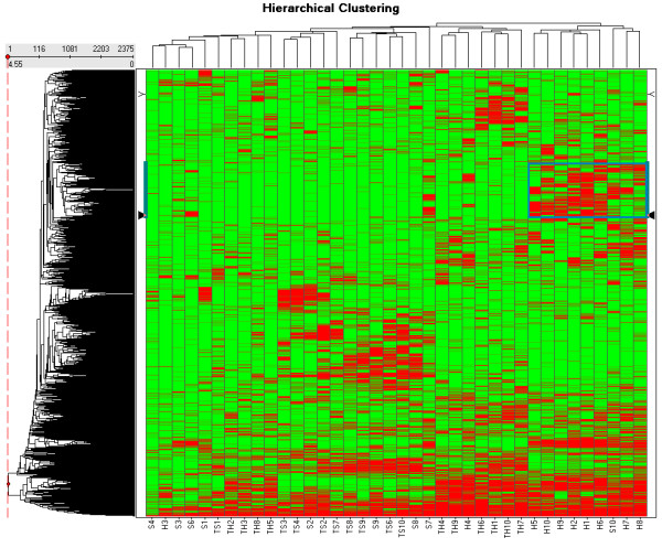
Figure 3.

Heat-map of the hierarchical clustering on the presence-absence peptide mass profile matrix in two dimensions of the 38 samples and 2375 peptide masses. The data of the clustering table is added as an additional file 7. A number of 10 spectra of glioma blood vessels with codes H1 to H10, 10 spectra of tissue surrounding the glioma vessels with codes TH1 to TH10, 10 spectra of normal vessels with codes S1 to S10, and 10 spectra of tissue surrounding the normal vessels with codes TS1 to TS10 are included. Two "normal vessels" samples, S5 and TS5, were excluded because they could not be calibrated. The highlighted box in Figure 3 represents the hierarchical clustering order 490 with mass 1037.5355 Da to 789 with mass 1665.7891 Da as presented (see Additional file 7).
