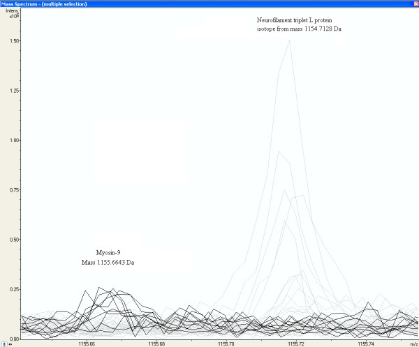
Figure 5.

Increased peak intensity at the mass MH+ 1155.6643 Da from a peptide of Myosin-9 in glioma vessel samples. The peaks of glioma vessel samples are represented by the dark lines in MALDI-FTICR mass spectra of all samples. The first isotopic mass of Neurofilament triplet L protein at 1154.7128 Da, represented by the grey lines, is as expected not present in Glioma vessels.
