Figure 2.
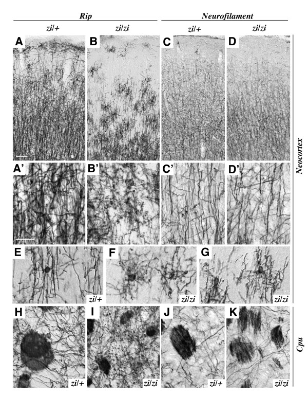
Developmental defect of oligodendrocytes in the zi/zi cerebrum. Coronal sections of the cerebral cortex (A-G) and caudate-putamen (H-K) at 4 weeks of age were immunostained with antibodies for Rip (A, B, E-I) or neurofilament (C, D, J, K). The genotype (zi/zi or control zi/+) is indicated in each panel. A-D, Cerebral cortex showing Rip-positive oligodendrocytes (A, B) and neurofilament-positive axons (C, D). The pial surface is at the top. A'-D', High-power photomicrographs of individual processes of Rip-positive oligodendrocytes (A', B') and neurofilament-positive axons (C', D') in the gray matter (layers II-III). In the zi/+ cortex, immunoreactive cells for Rip, which possessed relatively thick and branched processes, were abundantly distributed in cortical layers II-VI, but were sparse in superficial molecular layer I. By contrast, Rip-positive cells in the zi/zi cortex were significantly decreased in number throughout the cortex. E-G, High-power views of individual Rip-positive cell in layers I-II. Rip-positive cells in zi/+ rats represent the typical morphology of myelinating oligodendrocytes, with round cell bodies and branched processes aligned in parallel (E), whereas Rip-positive cells in zi/zi rats frequently showed a morphological abnormality of distorted-shaped cell-bodies with fine and wavy processes that branched irregularly (F, G). H-K, Caudate-putamen of the basal ganglia immunostained for Rip (H, I) and neurofilament (J, K). In the zi/zi rat, Rip-positive oligodendrocytes exhibited an abnormal morphology with thinner and arborized processes, frequently accompanied by many spherical or granule-like fragments (I). Extension of neurofilament-positive axons and axon bundles appeared normal in zi/zi rats (K). Scale bars: 50 μm (A-D); 20 μm (A'-D', E-K).

