Figure 3.
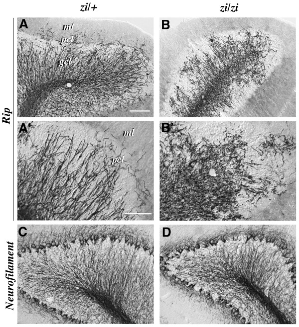
Developmental defect of oligodendrocytes in the zi/zi cerebellum. Sagittal sections of the cerebellum from zi/+ (A, A', C) or zi/zi (B, B', D) rats at 4 weeks of age were immunostained with antibodies for Rip (A-B') or neurofilament (C, D). A, B, Differentiating Rip-positive oligodendrocytes were distributed predominantly within the internal granule cell layer (gcl) and the white matter tracts in the zi/+ cerebellum (A), whereas Rip-positive processes of the zi/zi mutant cerebellum were distorted and irregularly arranged, and sparsely distributed within the gcl (B). A', B', Higher magnifications of the molecular layer (ml), Purkinje cell layer (pcl) and gcl shown in A and B, respectively. The cerebellar oligodendrocytes in zi/zi rats exhibited irregularly aligned short processes. C, D, The number, density and distribution pattern of neurofilament-positive axons in zi/zi cerebellar folia (D) were indistinguishable from those in the zi/+ cerebellar folia (C). Scale bars: 50 μm (A, B, C, D); 25 μm (A', B'). gcl, internal granule cell layer; ml, molecular layer; pcl, Purkinje cell layer.

