Figure 5.
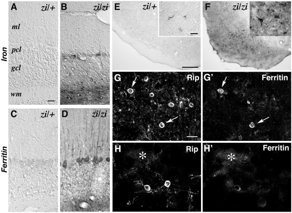
Up-regulation of ferritin in oligodendrocytes in zi/zi rats. A-D, Iron staining (A, B) or immunostaining of ferritin (C, D) in the cerebella of 8-month-old zi/zi (B, D) and age-matched zi/+ rats (A, C). E, F, Immunostaining of ferritin in the substantia nigra of 8-month-old zi/zi and zi/+ rats. Insets show ferritin-positive cells in the substantia nigra reticulata. G-H', Confocal images of double labeling for Rip and ferritin in the zi/zi substantia nigra, showing the increased expression of ferritin in Rip-positive oligodendrocytes (arrows). The diffuse immunoreactivity of ferritin was often observed around the Rip-positive oligodendrocytic processes (asterisk). Panels G/G' and H/H' represent pairs of double stained photomicrographs. Scale bars: 10 μm (A-D); 500 μm (E, F); 10 μm (insets in E, F); 10 μm (G-H'). wm, cerebellar white matter.

